

Leonardo da Vinci (1452-1519)
Recto: The vessels and nerves of the neck. Verso: The vessels of the liver c.1508
Pen and ink | 19.1 x 13.5 cm (sheet of paper) | RCIN 919051

Leonardo da Vinci (1452-1519)
Recto: The vessels and nerves of the neck. Verso: The vessels of the liver c.1508
Recto
©

Leonardo da Vinci (1452-1519)
Recto: The vessels and nerves of the neck. Verso: The vessels of the liver c.1508
-
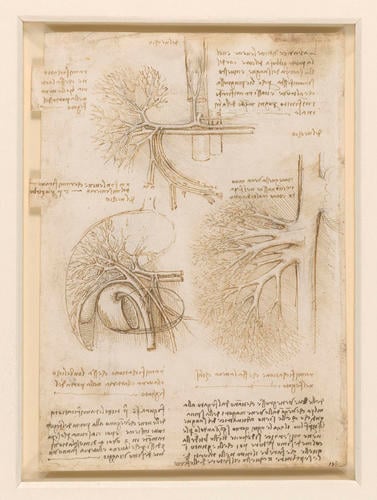
A folio from Leonardo's 'Anatomical Manuscript B'. All the drawings are labelled 'del vechio' ('of the old man'), and thus continue the series of dissections of the ‘centenarian’ (see 919027).
Recto: a drawing showing the front view of the blood vessels of the neck; a study of the phrenic or accessory phrenic nerve running between the artery and vein towards the heart; notes on the drawings.
Verso: the portal vein and branches of the coeliac artery; the portal vein and its branches; a detail of the liver, the gall bladder, the pylorus, the duodenum and blood vessels; notes on the drawings. In the drawing at centre left, an enlarged umbilical vein is seen curving diagonally from the liver to lower right: although the umbilical vein closes after birth (remaining vestigially as the round ligament of the liver), portal hypertension in the cirrhotic subject appears to have forced the vein open again.
In the upper drawing, the celiac axis is seen coming off the aorta (drawn incorrectly on the anatomical right of the torso) and splitting into the splenic and common hepatic arteries, drawn almost in a straight line; above their bifurcation is another branch, probably the left gastric artery. A smaller vessel branching off to the left (proper) from the celiac axis, almost at its junction with the aorta, could be the left phrenic artery or even an artery of the adrenal (suprarenal) artery. But the surrounding fascia of the aorta (tunica externa) is so thick in this area, and contains so many autonomic nerve fibres, that Leonardo’s dissection would have been severely hindered unless he had completely removed it to expose the tunica media of the artery.
Branching downwards from the common hepatic artery are the right gastric artery (labelled o at its cut end) and the gastroduodenal artery, which in turn branches into the superior pancreaticoduodenal (m) and right gastric-epiploic (p) arteries, all with venous corollaries. Directly below the splenic and common hepatic arteries, the hepatic portal vein is shown as continuous with the splenic vein, with no contribution from the superior mesenteric vein. The convolutions of the duodenum and the intertwining of the mesenteric and hepatic portal vessels continue to provide today’s students with difficulty, and faced with an extremely aged subject where atrophy or pathology could have complicated the anatomy, it is not surprising that Leonardo did not correctly illustrate this system.
In the drawing at centre left Leonardo has added the duodenum and outline of the stomach, and the gall bladder with its cystic duct merging with the hepatic duct to form the common bile duct. The bile duct is shown merging with the duodenum, but neither the pancreas nor the prominent junction of the pancreatic duct with the descending duodenum is indicated.The drawing to the right shows the formation of the hepatic veins, an upper and a lower group, draining separately into the upper end of the inferior vena cava, just below its termination in the right atrium of the heart (indicated in the margin of the sheet).
Text adapted from M. Clayton and R. Philo, Leonardo da Vinci: Anatomist, London 2012Provenance
Bequeathed to Francesco Melzi; from whose heirs purchased by Pompeo Leoni, c.1582-90; Thomas Howard, 14th Earl of Arundel, by 1630; probably acquired by Charles II; Royal Collection by 1690
-
Creator(s)
Acquirer(s)
-
Medium and techniques
Pen and ink
Measurements
19.1 x 13.5 cm (sheet of paper)
Other number(s)


