

Leonardo da Vinci (1452-1519)
Recto: Studies of the scapular, neck, and pelvic vessels. Verso: The vessels of the neck and shoulder c. 1508
Pen and ink over black chalk | 19.0 x 13.9 cm (sheet of paper) | RCIN 919049

Leonardo da Vinci (1452-1519)
Recto: Studies of the scapular, neck, and pelvic vessels. Verso: The vessels of the neck and shoulder c. 1508

Leonardo da Vinci (1452-1519)
Recto: Studies of the scapular, neck, and pelvic vessels. Verso: The vessels of the neck and shoulder c. 1508
-
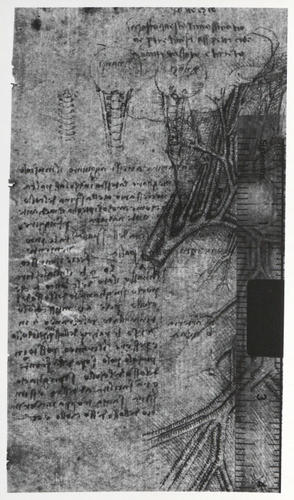
A folio from 'Leonardo's Anatomical Manuscript B'. Recto: a study of the right scapula (shoulder blade) and the long head of the biceps muscle seen from behind; three small diagrammatic studies, two showing the skull, spinal column and sterno-cleido mastoid, the third showing the spinal column only; and a diagram showing intercommunicating arteries in front and intercommunicating veins behind.
Verso: a study of the arteries and veins of the right side of the neck and shoulder of a man seen in profile. Leonardo labelled the drawing del vechio, and thus it purports to show the arteries and veins of the neck, shoulder and upper arm of the ‘centenarian’. Leonardo lettered the vessels and annotated the drawing thus:
"a are the ramifications of the artery
b is the ramification of the veins
c is the cephalic vein
n are the two vessels which enter the cervical vertebrae to nourish them
o is the basilic vein
s are the apoplectic vessels"
The cut artery a is thus the aorta: its branching is symmetrically arranged (following a bovine rather than human pattern), giving rise to both right and left common carotid and right subclavian arteries – the left subclavian is not visible in this drawing but its presence can be inferred. The superior vena cava, b, is formed from the union of left and right brachiocephalic veins. The vertebral arteries n are a long way out of position, placed too high in the neck. The common carotid artery and internal jugular vein are the straight vessels passing to the right of s; to the left of s are the external jugular vein (receiving a branch from the posterior external jugular, running around the back of the neck) and an accompanying artery. In reality there is no artery that truly accompanies the external jugular vein – the only candidate is the thyrocervical trunk and its branches – but Leonardo believed that every vein was accompanied by a similar calibre artery. The arterial channel from subclavian to axillary to brachial is shown, along with the corresponding veins.
The note at upper left states:
"If you close the four vessels at m on each side of the throat, he who has them closed will fall to the ground immediately in sleep as if dead, and he will never wake up on his own. And if he is left for one-hundredth of an hour in such a condition he will never wake again, neither on his own nor with the help of others."
This describes the long-known phenomenon (described, for example, by Mondino) that pressure in this area can lead to unconsciousness. Rather than pressure on the vessels themselves, this is due to pressure on the baroreceptors of the carotid sinus, as had in effect been noted in antiquity: the Greek physician Rufus of Ephesus (c. AD 100) wrote in his treatise On the names of parts of the human body that ‘the ancients called the arteries of the neck carotid [Greek κάρωτιδες, ‘stupefying vessels’], because they believed that when they were pressed hard, the animal became sleepy and lost its voice; but in our age it has been discovered that this accident does not proceed from pressing upon these arteries, but upon the nerves contiguous to them’.
Text adapted from M. Clayton and R. Philo, Leonardo da Vinci: Anatomist, London 2012.Provenance
Bequeathed to Francesco Melzi; from whose heirs purchased by Pompeo Leoni, c.1582-90; Thomas Howard, 14th Earl of Arundel, by 1630; Probably acquired by Charles II; Royal Collection by 1690
-
Creator(s)
Acquirer(s)
-
Medium and techniques
Pen and ink over black chalk
Measurements
19.0 x 13.9 cm (sheet of paper)
Other number(s)


