Recto: The stomach and related structures. Verso: The abdomen c.1508
Recto: Pen and ink over traces of black chalk. Verso: Pen and ink over black chalk | 19.2 x 14.1 cm (sheet of paper) | RCIN 919039
-
A folio from Leonardo's 'Anatomical Manuscript B'.
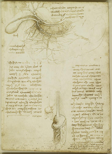
Recto: a study of the gall bladder and related blood vessels; two drawings of the oesophagus, stomach and greater omentum, one on a smaller scale; notes on veins and arteries and their alteration in old age.
The principal drawing (labelled 'del vechio', 'of the old man'; see 919027) shows details of the anatomy adjacent to the stomach and liver. The stomach is lightly outlined below and to the right of the drawing, and the liver is lifted away so that we are looking into the region between the two organs. The area of diagonal shading at the centre of the drawing corresponds with the lesser omentum, a clear membrane between the liver and the stomach and first duodenal segment. The gallbladder is to the left, with its duct and the cystic artery. The splenic and common hepatic arteries are drawn as if they were one continuous artery (labelled c a b), with the splenic and hepatic portal vein immediately below and partially obscured. The left and right gastric arteries branch off from the common hepatic artery at b and a respectively, their ramifications meeting on the lesser curvature of the stomach, with further branches spreading out over the surface of the stomach. The gastroduodenal artery should also begin around a, travelling behind the duodenum; possibly Leonardo has attempted to show the common hepatic artery trifurcating at that point, but the details are impossible to resolve.
Leonardo appears to have been confused by the various elements of the peritoneum, the membrane that forms the lining of the abdomino-pelvic cavity. In the larger drawing below, showing the stomach from the right, he correctly depicts the greater omentum attached to the stomach. The membrane apparently shown hanging down (shaded) behind the duodenum and first portion of small intestine is, however, incorrect – unless in the process of dissection, a part of the peritoneum came loose from the posterior body wall. In the smallest, sketchiest drawing, Leonardo seems to have illustrated the transverse mesocolon (the supporting mesentery of the transverse colon and part of the stomach) attached to the greater omentum, though lifted up; in its normal position it lies just below the stomach.
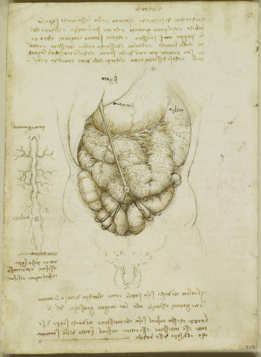
Verso: a study of the abdominal organs, i.e. liver, spleen. stomach and intestines, and another of the blood vessels of the anterior abdominal wall; notes on the drawings.
The main drawing shows the abdomen from the front with the abdominal wall and peritoneum removed – Leonardo called these structures mirach and sifac respectively, the terms used by Leonardo’s principal source Mondino and derived from the Arabic of Avicenna and his peers. We see the liver (labelled fegato), stomach (stomaco) and spleen (milza) labelled above, with the membrane of the greater omentum rendered almost transparent and covering the small intestine. A note reads: ‘the net [omentum] which lies between the sifac [parietal peritoneum] and the intestines in the old covers all the intestines and is drawn back between the bottom of the stomach and the upper part of the bowel [transverse colon]’.
Although Leonardo does not state that this depiction resulted from the dissection of the ‘centenarian’, the enlarged spleen and the presence of the umbilical vein indicate that it was based on that cirrhotic subject. Leonardo has also illustrated, below the umbilicus, two paired remnants of the umbilical arteries (the medial umbilical ligaments) wrapping around the colon – actually there is only one such structure on each side. Just to the anatomical left of the umbilicus is a large loop of what appears to be transverse colon. Below Leonardo wrote ‘the colon in the old becomes as slender as the middle finger of the hand, and in the young it is like the greatest thickness of the arm’, but the intestines appear to be inflated with gas, as would happen as part of the putrefaction process.In the drawing to the left, superior and inferior epigastric vessels (whether arteries or veins is unclear) are shown joining or anastomosing around the umbilicus. At the top of that drawing is the xiphoid process, at the end of the sternum, with a bifurcated form (a natural variation) and labelled with the picturesque term pomogranato.
Text from M. Clayton and R. Philo, Leonardo da Vinci: Anatomist, London 2012Provenance
Bequeathed to Francesco Melzi; from whose heirs purchased by Pompeo Leoni, c.1582-90; Thomas Howard, 14th Earl of Arundel, by 1630; Probably acquired by Charles II; Royal Collection by 1690
-
Creator(s)
Acquirer(s)
-
Medium and techniques
Recto: Pen and ink over traces of black chalk. Verso: Pen and ink over black chalk
Measurements
19.2 x 14.1 cm (sheet of paper)
Other number(s)


