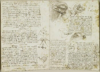
The right ventricle and the valves of the heart (recto); The ventricles, valves and papillary muscles (verso) c. 1511-13
Pen and ink | 22.1 x 31.2 cm (sheet of paper) | RCIN 919118
-
Recto: On the left half of the double page (historically RL 19118r), notes on the pulmonary valves of the heart, with three sketches; coronary vessels seen from apex of heart. On the right half (RL 19119r), three semi-schematic drawings and two diagrams of the ventricles of the heart, with notes on their action. The sheet was compiled by Leonardo as two distinct folios, though there are no stitch-marks down the central fold to suggest it was ever bound up.
The three largest drawings on the right-hand page show the interior of the right ventricle from below, with the moderator band and two of the three papillary muscles prominent, and the chordae tendineae (particularly well drawn) extending up to the tightly closed cusps of the tricuspid valve. The geometrical diagram to the centre of the page purports to show the location of the papillary muscles on the walls of the ventricle; again only two are indicated. The moderator band is highlighted in the schematic view of the heart below. Leonardo referred to this band of tissue, extending between the interventricular septum and the base of the anterior papillary muscle, as ‘the chain of the right ventricle’, and until relatively recently it was thought to prevent overexpansion of the right ventricle; it is now known to play a role in the electrical conduction system of the heart.
On the left-hand page Leonardo considers the function of the pulmonary valve, which lies between the right ventricle and the pulmonary trunk. The first of the marginal drawings is a partly geometrical rendering of the valve: the semilunar attachments of the cusps are well indicated, with sketched lines on the uppermost cusp presumably representing the chordae. In a note Leonardo suggests that the curved, fleshy attachment of the cusps was designed to deflect the ‘impetus’ of the blood as it flows into the cusps when the valve closes, otherwise the ‘impetus’ would damage the cusps. Leonardo was increasingly concerned that his investigations should have a rational basis: scrawled in the top margin is the exhortation ‘let no-one who is not a mathematician read my principles’.
The next marginal sketch is a cross-section of the aortic valve, showing the swirling of the blood in the sinus of Valsalva (studied exhaustively on 919082-3, 919116 etc). Next is a view of the base of the heart from above, rendered as a rounded triangle with the aortic valve at the centre (‘in the principal position at the base of the heart, just as it holds principal place in the life of the animal’). Lastly is a sketch of the heart viewed from its apex, the right side to the top, with the anterior and posterior interventricular arteries meeting near the apex, as described in the accompanying note.
*
Verso: On the left half of the double page (historically RL 19119v), two drawings of a section of the right ventricle of the heart, with notes on its action; above, a light preliminary sketch of a dissection of the heart; top right, a small drawing and diagram of a ventricle of the heart. On the right half (RL 19118v), five studies of the ventricle of the heart, seen from the side and from underneath; two skeletal studies of the right arm, demonstrating pronation & supination, with notes.
The sketches to upper right show the base of the bovine heart sectioned through, with the atria reflected to either side (represented by rough looping lines). The right ventricle is the crescent-shaped cavity at the top of each image, the left ventricle is the irregular triangle to lower right, the aortic valve is at the centre, and the pulmonary valve is to the left (Leonardo labels the pulmonary artery as the ‘venous artery’). Adjacent to those sketches is a section through the left ventricle, with the aortic valve at top left; though Leonardo has shown papillary muscles and the chordae tendineae reaching up to the rim of the ventricle, the mitral valve is not really present.
The drawings towards the bottom of the left-hand page examine the interior of the right ventricle. That on the right imagines the triscuspid valve distended to give a view down into the ventricle: the chordae tendineae are shown arising from the papillary muscles and attaching most of the way around this aperture, with the moderator band dimly seen passing from centre left to lower right. The study at lower left is a section of the right ventricle at the level of the moderator band, with its two attachments labelled ‘A’. The view is now upwards towards the closed tricuspid valve. Leonardo was still uncertain about the correct number of papillary muscles (three in the right ventricle, two in the left) – in this drawing he depicts five, in the previous drawing four, and on the other side of the sheet, two. In the right ventricle, the posterior and septal papillary muscles in particular can appear to be a small cluster of muscles. In 919078 he established the correct number in each ventricle.
The notes and sketches at upper left examine the spatial relationship between the ventricles and the superficial vessels of the heart. A further note seeks confirmation of Leonardo’s beliefs about the source of heat in the heart:
Observe whether the revolution of milk when butter is made, heats it. And by such means you will be able to test the power of the auricles [atria] of the heart, which receive and expel the blood from their cavities ... these being made only in order to heat and refine the blood and make it more quickly penetrate the wall through which it passes from the right to the left ventricle.
At lower right is yet another study of the pronation and supination of the forearm, an action that Leonardo had analysed extensively a couple of years earlier in Manuscript A (919000v, 919004r) and that continued to fascinate him – similar studies are found on an embryological sheet, 919103v.Provenance
Bequeathed to Francesco Melzi; from whose heirs purchased by Pompeo Leoni, c.1582-90; Thomas Howard, 14th Earl of Arundel, by 1630; Probably acquired by Charles II; Royal Collection by 1690
-
Creator(s)
Acquirer(s)
-
Medium and techniques
Pen and ink
Measurements
22.1 x 31.2 cm (sheet of paper)
Other number(s)
RL 19119


