

The rotation of the arm, and the foetus in the womb c.1511
Pen and ink over black chalk | 28.7 x 21.1 cm (sheet of paper) | RCIN 919103
-
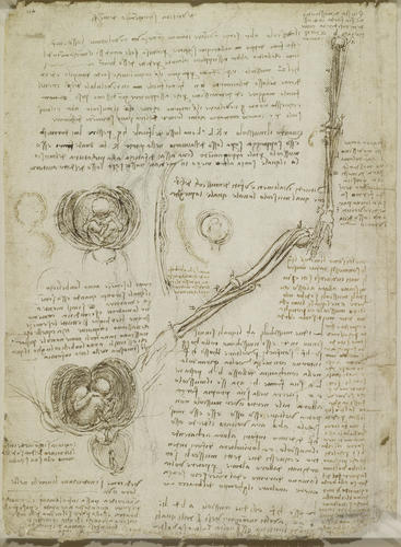
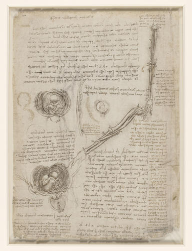
Studies of the bones of the right arm, and notes on the action of the muscles; studies of a fetus in utero; sketches of the uterine wall and cotyledons of a cow's placenta.
At centre and upper right are two studies of the arm, concentrating on the rotation of the hand and forearm (cf. RCIN 919000v, 919004r). Both drawings show biceps, originating from two heads on the scapula, inserting on the upper forearm, and responsible in part for supination (turning the arm so that the palm faces upwards); and pronator teres, originating mainly on the humerus with a secondary head on the ulna (as described by Leonardo in the accompanying notes), inserting on the radius and responsible for pronation. The muscles are reduced in thickness, and in the diagram at upper right they are further reduced to threads, with brachialis also shown along the left margin of the humerus. While brachialis can flex the arm regardless of the position of the forearm, biceps can only flex the arm when it is supinated, and thus it will supinate a pronated forearm when it acts.
This subtle understanding dates the sheet soon after completion of Leonardo's 'Anatomical Manuscript A' – the drawing at top right is essentially a continuation of those on 919004r. During this period Leonardo’s attention would shift away from the mechanical aspects of the muscles and bones to the mysteries of the heart and reproduction. The small sketch at the centre of the page shows the fetus surrounded by its membranes; in the larger drawings to the left these membranes are peeled away and opened out, and in that at lower left the vagina is similarly sectioned, with an ovary and the uterine ligaments sketched to the left. Most of the drawings show the multiple, cotyledenous placenta derived from bovine dissection (see 919055r) but found throughout Leonardo’s supposedly human embryological studies of this period. The details at centre and centre left investigate (in cross-section) whether these cotyledons bulge outwards, inwards or both.
Leonardo was acutely conscious of the universality of reproduction. It is not fanciful to see botanical aspects in his drawings of the fetus in the womb, echoes of unfurling flowers or an opening nut, for in his note at centre left here he writes:
"All seeds have an umbilical cord which is broken when the seed is ripe. Likewise they have a uterus and membranes, as herbs and all seeds that are produced in pods demonstrate. But those which are produced in nutshells, such as hazelnuts and pistachios, have a long umbilical cord which shows itself in infancy."
(The recto of the sheet, as specified in the Windsor inventory, is blank; all the drawings are on the verso.)
Text from M. Clayton and R. Philo, Leonardo da Vinci: Anatomist, London 2012
Provenance
Bequeathed to Francesco Melzi; from whose heirs purchased by Pompeo Leoni, c.1582-90; Thomas Howard, 14th Earl of Arundel, by 1630; Probably acquired by Charles II; Royal Collection by 1690
-
Creator(s)
Acquirer(s)
-
Medium and techniques
Pen and ink over black chalk
Measurements
28.7 x 21.1 cm (sheet of paper)
Other number(s)


