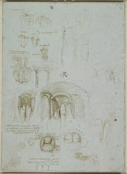
The left ventricle and mitral valve c.1512-13
Pen and ink on blue paper | 28.4 x 20.9 cm (sheet of paper) | RCIN 919080
-
Studies, some architecturally stylized, of the heart, its valves, atria and ventricular orifices; notes on the drawings. On the verso, a black chalk sketch of a human figure running to the left.
At top left are two rough sketches of the heart with the coronary and other superficial vessels. Below are three sections of the heart – a schematic longitudinal section with the atria and ventricles shaded, and two transverse sections, one at the approximate level of the valves to show (inaccurately) their relative positions, another lower down cutting through the ventricles. To the left of these is an outline of the heart with two vertical lines indicating the sections that would each cut through one ventricle only. The results of this are shown in the two sketches below, in which the heart is cut into three portions and opened out, thus exposing the inner surfaces of each ventricle.
The two largest studies at centre and centre left present details of the interior of the left ventricle. We see the two papillary muscles arising in the lower part of the ventricle, with the chordae tendineae connected to the cusps of the mitral valve. Below, the mitral valve is shown in closure, from the atrial side. The mitral valve is normally regarded as two-cusped, and indeed its alternative name is the bicuspid valve; but Leonardo has accurately shown, on either side of the major cusps, two smaller commissural cusps, which are relatively larger in the mitral valve of the ox and other large mammals than in the human.
A pale sketch at bottom right shows the four valves of the heart (which lie in approximately the same plane) from above; just below the mitral and aortic valves Leonardo has written osso, referring to the two small bones that lie in the fibrous aortic ring of the bovine heart. In that drawing the mitral and tricuspid valves are closed and the aortic and pulmonary valves are open. This is the configuration during the second phase of systole, when the ventricles contract to pump blood into the aorta and pulmonary artery. If Leonardo did intend to display this configuration, rather than showing the valves arbitrarily open or closed, then this is an astonishing piece of observation and implies a very clear understanding of the sequence of operation of the heart.
Text from M. Clayton and R. Philo, Leonardo da Vinci: Anatomist, London 2012
Provenance
Bequeathed to Francesco Melzi; from whose heirs purchased by Pompeo Leoni, c.1582-90; Thomas Howard, 14th Earl of Arundel, by 1630; Probably acquired by Charles II; Royal Collection by 1690
-
Creator(s)
Acquirer(s)
-
Medium and techniques
Pen and ink on blue paper
Measurements
28.4 x 20.9 cm (sheet of paper)
Object type(s)
Other number(s)


