Recto: The cranium sectioned. Verso: The skull sectioned 1489
Recto: Pen and ink. Verso: Traces of black chalk, pen and ink | 19.0 x 13.7 cm (sheet of paper) | RCIN 919058
-
For Leonardo's skull studies of 1489, see RCIN 919059.
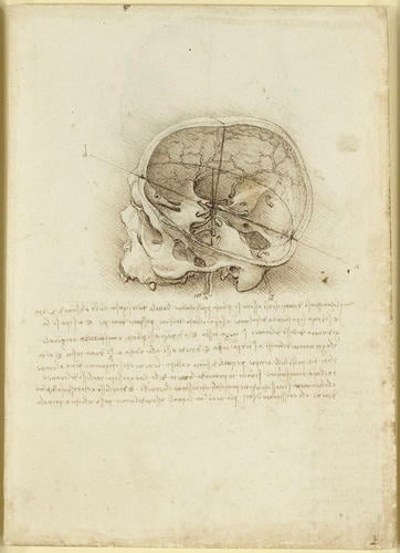
The cranium studied on the recto is in the same section as in the upper drawing of 919057r, with the floor of the cranial cavity tilted a little further towards the viewer. Again, the site of the senso comune is indicated at the intersection of orthogonal axes. The note defines this position in proportional terms:
The confluence of all the senses has below it in a perpendicular line the uvula, where one tastes food, at a distance of two fingers; and it is directly above the windpipe of the lung and the orifice of the heart by a space of one foot. And it has the junction of the bones of the head half a head above it; and in front of it in a horizontal line is the lacrimator of the eye at one-third of a head.
Leonardo was in all probability working with a dry skull, with the dura mater absent, and he attempted to infer the pathways of some of the nerves and vessels, with variable success. He drew the mastoid emissary vein n passing through the mastoid foramen, with its pair on the other side of the cranium, though he does not indicate their origin or destination. Two vessels labelled a and m travel to the middle of the cranium from below: a is probably the middle meningeal artery, seen entering the cranium through the foramen spinosum; m enters the cranium through the foramen ovale, where it then turns through 90º and travels forwards to the floor of the orbit. That should be the infraorbital artery, which arises from the maxillary artery; but the artery that passes through the foramen ovale is the accessory meningeal, a separate branch of the maxillary artery.
Around the nose are the angular and dorsal nasal veins, with their connections to the frontal (supratrochlear) vein. Further vessels are shown ramifying on the inside of the frontal and parietal bones, and Leonardo discusses how these are partially embedded in the bone and covered by the meninges: ‘The veins [they are in fact arteries] which are drawn within the cranium proceed in their ramification, imprinting half their thickness into the bones of the cranium, and the other half is hidden within the membranes which cover the brain.’
The optic nerves can be seen emerging from the optic foramina and converging on the supposed site of the senso comune. Immediately below are two further structures: the first is probably the oculomotor nerve (CNIII), with its pair just visible on the other side of the optic chiasm; the second is probably the maxillary (or possibly the ophthalmic) division of the trigeminal nerve (CNV; the mandibular division, which should pass downwards through the foramen ovale, is not shown).*
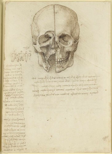
In the drawing on the verso of the sheet, Leonardo first sawed the skull in the median plane, dividing it into equal left and right halves; then he sawed away the front of the right side of the skull – a very difficult operation, given the delicacy of the bones between the facial sinuses. By juxtaposing these two halves, the viewer is able to locate the cavities of the skull in relation to the surface features; covering the left (proper) side of the skull dramatically reduces the legibility of the demonstration.
The sectioned right side shows the frontal sinuses (at the level of the eyebrows) and the opening into their drainage duct. In the orbit of the eye can be seen the superior and inferior orbital fissures, the nasolacrimal canal leading into the inferior meatus of the nasal cavity, the infraorbital groove, and on the cut margin, the infraorbital canal. Below the orbit is the maxillary sinus and its opening into the nasal cavity. Two teeth are shown sectioned, with their roots. In the mandible or jawbone can be seen the mandibular canal and the mental foramen (the small hole connecting the mandibular canal with the surface of the mandible), and on the inside of the mandible is the profile of the mylohyoid line. On the intact left side of the skull are the supraorbital notch, the fossa for the lacrimal sac on rthe intact orbital rim, the canine fossa, the oblique line of the mandible, and the mental foramen.
In his note Leonardo relates the site of the senso comune to these cavities:
The cavity of the eye socket, and the cavity of the bone that supports the cheek, and those of the nose and mouth, are of equal depth and terminate in a perpendicular line below the senso comune. And each of these cavities is as deep as the third part of a man’s face, from the chin to the hair.
In the left margin Leonardo added stylised drawings of each of the different types of teeth—molar, premolar, canine and incisor – counting them in the note below to a total of 32, including the last molars or ‘wisdom teeth’. While the correctness of this figure may seem trivial, there was no consensus at the time (probably due to the sporadic eruption of wisdom teeth), with some commentators repeating Aristotle’s assertion that women have fewer teeth than men.
Text adapted from M. Clayton and R. Philo, Leonardo da Vinci: Anatomist, 2012, no. 13.Provenance
Bequeathed to Francesco Melzi; from whose heirs purchased by Pompeo Leoni, c.1582-90; Thomas Howard, 14th Earl of Arundel, by 1630; probably acquired by Charles II; Royal Collection by 1690
-
Creator(s)
Acquirer(s)
-
Medium and techniques
Recto: Pen and ink. Verso: Traces of black chalk, pen and ink
Measurements
19.0 x 13.7 cm (sheet of paper)
Markings
watermark: Crown, partial [-]
Other number(s)


